Showing posts with label Guest Bloggers. Show all posts

Saturday, March 24, 2012

Updated: Thyroid Cancer Staging Guide


The stage of a cancer is a description (usually numbers I to IV with IV having more progression) of the extent the cancer has spread. The stage often takes into account the size of a tumor, how deeply it has penetrated, whether it has invaded adjacent organs, how many lymph nodes it has metastasized to (if any), and whether it has spread to distant organs. Staging of cancer is the most important predictor of survival, and cancer treatment is primarily determined by staging. Thus, staging does not change with progression of the disease as it is used to assess prognosis. 
A patients' cancer, however, may be re-staged after treatment but the staging established at diagnosis is rarely changed. Cancer staging can be divided into a clinical stage and a pathologic stage. In the TNM (Tumor, Node, Metastasis) system, clinical stage and pathologic stage are denoted by a small "c" or "p" before the stage (e.g., cT3N1M0 or pT2N0).
Because they use different criteria, clinical stage and pathologic stage often differ. Pathologic staging is usually considered the "better" or "truer" stage because it allows direct examination of the tumor and its spread, contrasted with clinical staging which is limited by the fact that the information is obtained by making indirect observations at a tumor which is still in the body. 
However, clinical staging and pathologic staging should complement each other. Not every tumor is treated surgically, therefore pathologic staging is not always available. Also, sometimes surgery is preceded by other treatments such as chemotherapy and radiation therapy which shrink the tumor, so the pathologic stage may underestimate the true stage. This staging system is used for most forms of cancer, except brain tumors and hematological malignancies
The American Joint Committee on Cancer (AJCC) created the following staging system for Thyroid Cancer Staging
  • T1 - Tumor diameter 2 cm or smaller
  • T2 - Primary tumor diameter greater than 2-4 cm
  • T3 - Primary tumor diameter greater than 4 cm limited to the thyroid or with minimal extrathyroidal extension
  • T4a - Tumor of any size extending beyond the thyroid capsule to invade subcutaneous soft tissues, larynx, trachea, esophagus, or recurrent laryngeal nerve
  • T4b - Tumor invades prevertebral fascia or encases carotid artery or mediastinal vessels
  • TX - Primary tumor size unknown, but without extrathyroidal invasion
  • NO - No metastatic nodes
  • N1a - Metastases to level VI (pretracheal, paratracheal, and prelaryngeal/Delphian lymph nodes)
  • N1b - Metastasis to unilateral, bilateral, contralateral cervical, or superior mediastinal mode metastases
  • NX - Nodes not assessed at surgery
  • MO - No distant metastases
  • M1 - Distant metastases
  • MX - Distant metastases not assessed
Stage I (any T, any N, M0)
Stage II (any T, any N, M1)


Author: Mark E Gerber, MD, FACS, FAAP  Clinical Assistant Professor of Otolaryngology, University of Chicago, Pritzker School of Medicine; Section Head, Pediatric Otolaryngology-Head and Neck Surgery, NorthShore University HealthSystem  


Co-Author: Brian Kip Reilly, MD  Assistant Professor of Otolaryngology and Pediatrics, Department of Otolaryngology, Children's National Medical Center, George Washington University School of Medicine

Friday, September 30, 2011

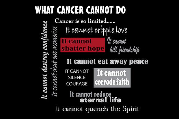
What Cancer Cannot Do


As soon as I read ”What Cancer Cannot Do” I felt my inner cancer SURVIVOR come out.  We tend to give cancer a lot of power.  It is part of the process to start blaming changes in our lives, relationships, outlook, attitude and choices on cancer.  The real deal is that each one of us has the power and inner-strength to focus on what we CAN do and what cancer CANNOT do.  It is a powerful exercise. 
I started thinking about what else could be added to the list.  I’m sharing it with all of my blog readers and asking you to add to the list as well.  Here is my version:
Cancer is so limited…
It cannot turn my world to darkness.
It cannot make the beauty of nature disappear.
It cannot take over my being.
It cannot drain my creativity.
It cannot silence my voice.
It cannot prevent me from spreading awareness and hope.
It cannot come between my connection with a Higher Power.
It cannot stop me from laughing and smiling.
It cannot take my will power.
It cannot make me give up.
It cannot take away my joy.
It cannot change my purpose in life.
ABOUT THE AUTHOR: 
Jennifer Bridge is a thyroid cancer survivor and Life Coach.  Jennifer offers complimentary intro life coaching sessions via phone. For details on how to schedule your complimentary coaching call please contact her at freelifecoachingcall@jenniferbridge.com 

Wednesday, August 24, 2011

Cultivating Gratitude

Feeling grateful can be a calming, serene and spiritual way of life, but for people on the cancer journey, gratitude can be -undestandably - a difficul concept.

Gratitude isn't a new idea. Most spiritual traditions emphasize the value and importance of having compassion for others. In the past several decade there has been a shift to look at gratitude as a useful tool rather and an idea.

Practicing gratitude means appreciating what you have and focusing on the support you receive from others.

With regular practice, gratitude, often can reduce anger and blame, while increasing joy and peace of mind. Being in a grateful state does not mean being blindly optimistic or ignoring unpleasant or negative things. It does involve, however, a shift in the thought process to look at things through a positive lens. 

Can gratefulness become a part of your life during the cancer journey?

Yes! However, it takes thought and intention to practice gratitude. Gratefulness can be another way to cope during the cancer journey. One ritual that promotes feeling grateful is writing a word or sentence describing what you feel grateful for and placing it in a "grateful" jar or box every day. This can help you put life in perspective. When you need a "gratitude" boost, dip into the jar and feast.

Here are few more simple and easy ways to beging practicing gratitude:

  • Write a thank-you note -- it feels good to make others happy. 
  • Work in a garden -- it is almost magical to see flowers grow.
  • Walk barefoot on a beach -- the ocean waves wash away fears.
  • Listen to music -- let the sound take you away.
  • Enjoy nature -- everything under the sun is the gift of life.
  • Tell someone you love them -- we know it but the sound of those words are very powerful and reassuring.

Recently, Eileen, a 46 year old cancer survivor, shared and experience of walking, deep in thought, gazing down at the sidewalk. She happened to glance up and see a big, beautiful maple tree in it's autum splendor. The tree reminded her of all the beauty of the world. As she looked higher, she noticed tree tops formed a circle over her head and opened up to the deep blue sky to reveal a couple of big puffy white clouds.

Following that experience, Eilieen noted," I felt really uplifted and grateful for the moment and all the little blessings I've seen since I first felt my lump. I couldn't help but feel close to my [deceased] mom and dad because I know they are there for me. I told them to hold hands form a big prayer circle and get to work! When I got home and turned up the driveway.  I saw a huge rainbow arching all the way accross the treetops in my backyard, full of color and bright with promise."

Rather than continuing to look down, Eileen literally changed her focus and looked up. Her inward, focus shifted from fear to being able to feel grateful -- for nature and for the support of those who loved her and whom she loved.  When you are grateful, you can become more optimistic, energetic, joyful, better equipped to handle challenges and more likely to help others. Additional benefits include closer ties to family and friends and a deeper sense of purpose.

A grateful heart is part of experiencing a full life -- one where you feel energized to reach out, help others, and have the power to make positive changes in your personal life and in your community. Try it for yourself and see what changes occur.

Editors Note: Article first published by Coping Magazine February 2008 Mary Bornstein is a program staff member at The Gathering Place, a cancer support center located in northeast Ohio. Betsy Kohn is the organization's director of volunteers. They have developed gratitude workshops to help individuals and families find additional ways of coping while on the cancer journey.

To reach Mary or Betsy call (216) 595-9546 or you can  
email   Mary   bornstein@touchedbycancer.org
email   Betsy kohn@touchedbycancer.org

Saturday, April 9, 2011

New radiation treatment practice recommendations for thyroid disease

New recommendations from the American Thyroid Association (ATA) on outpatient radioiodine (131I) treatment aim to minimize unintended radiation exposure and maximize the safety of patients, their families, and the public. The new ATA recommendations are presented in the April issue of Thyroid, a peer-reviewed journal published by Mary Ann Liebert, Inc
The ATA convened a task force to update radiation safety information related to outpatient 131I therapy to treat  and . The new ATA practice recommendations cover a broad range of topics including travel; safety precautions at home, work, and school; personal hygiene; and pregnancy and breastfeeding. 
These recommendations comply with the most up-to-date U.S. Nuclear Regulatory Commission (NRC) regulations, including a recent guidance statement that advises medical professionals administering 131I therapy to ask patients about their intended destination after the treatment and to discourage them from staying at hotels to limit public .
In a Commentary in the February issue of Thyroid, Richard T. Kloos, MD, Professor, The Ohio State University and Secretary/Chief Operating Officer of the ATA, states that the new ATA document "aims to provide simplified, consistent, and safe instructions for care providers and patients."
"The strength of these practice recommendations is that the task force included representatives across the range of disciplines that use radiation to treat thyroid patients. It is essential that our patients receive clear and consistent information from those ordering, administering, and monitoring these treatments," states Gregory A. Brent, MD, Professor of Medicine and Physiology, David Geffen School of Medicine at the University of California Los Angeles and President of the ATA.
More information: The ATA recommendations are available free online at  www.liebertpub.com/thy
Provided by Mary Ann Liebert, Inc

Friday, April 8, 2011

Diagnosing Pediatric Thyroid Cancer: Biopsy

  • FNAB is the criterion standard in the diagnostic workup of adult thyroid nodules. Several studies report efficacy in the pediatric population.
  • High diagnostic accuracy with experienced pathologists improves the selection of pediatric patients for surgery and is an adjunct to guide further management.
  • Ultrasonography can be a useful guide for percutaneous needle biopsy when the lesion is difficult to identify with palpation.
  • FNAB is often not practical in children younger than 10 years; therefore, excisional biopsy (surgical removal of nodule or tumor mass) under general anesthesia is recommended in this population.
  • Using molecular polymerase chain reaction (PCR) studies on FNAB aspirate is mostly beneficial in the clinical research setting. It can be used in a very small number of patients for diagnostic purposes, but it remains expensive.

    Significant Histologic Findings Review


    Follicular adenoma is the most common cause of solitary nodules of the thyroid in the pediatric population. Adenomas are solitary, well circumscribed, and well encapsulated and are composed of glandular epithelium. Most are histologically follicular but are occasionally papillary.
    • Most thyroid cancers (papillary, follicular, anaplastic) originate from follicular cells. Medullary thyroid cancers (25% hereditary vs 75% sporadic) are of C-cell (calcitonin-producing) origin.
    • Thyroid malignancies in children are usually well-differentiated papillary or papillary-follicular subtypes, but all histologic types have been observed. 
    • Papillary carcinoma lesions, which comprise an estimated 72% of pediatric thyroid cancers, are irregular, solid, or cystic masses that arise from follicular epithelium.
    • Microscopically, pediatric thyroid cancer masses or nodules have fronds of epithelium and distinct uniform cells with rare mitoses. Most contain both papillary and follicular components. The cells contain pink, finely granular cytoplasm with large pale nuclei (Orphan Annie eyes) and nuclear grooves. 
    • Psammoma bodies (rounded calcified deposits) can be found in approximately 50% of the lesions. 
    • Pediatric Papillary Carcinoma has frequent lymphatic and pulmonary metastases.

    • Follicular carcinoma lesions, which comprise 18% of pediatric thyroid cancers, are usually encapsulated and have highly cellular follicles and microfollicles with compact dark-staining nuclei of fairly uniform size, shape, and location. Pathologic diagnosis can be made only when invasion of the capsule, adjacent glands, lymphatics, or blood vessels is seen. 
    • Pediatric Follicular Carcinoma metastasizes intravascularly to the lungs, brain, and bones. When a portion of the cells in the tumor are found to be oxyphilic (Hürthle cells), it is called a Hürthle cell tumor. These lesions tend to have a less favorable prognosis.
    • MTC or Pediatric Medullary Thyroid Cancer arises from the thyroid parafollicular or C cells, which secrete calcitonin and are derived from the neural crest and ultimobranchial body. Hyperplasia of the C cells is thought to represent a precancerous state. 
    • Histologically, MTC is composed of columns of epithelial cells and dense stroma that typically stain for amyloid and collagen. The nuclei are hyperchromatic, and mitoses are common. The cells have a fusiform shape and may form a whirling pattern. Calcifications are observed in 50% of these lesions.
    Author: Mark E Gerber, MD, FACS, FAAP  Clinical Assistant Professor of Otolaryngology, University of Chicago, Pritzker School of Medicine; Section Head, Pediatric Otolaryngology-Head and Neck Surgery, NorthShore University HealthSystem  

    Co-Author: Brian Kip Reilly, MD  Assistant Professor of Otolaryngology and Pediatrics, Department of Otolaryngology, Children's National Medical Center, George Washington University School of Medicine 
    Thursday, April 7, 2011

    Diagnosing Pediatric Thyroid Cancer: Imaging Studies

    Imaging studies reveal the malignant potential and the extent of disease, and they provide an anatomical roadmap for surgical planning. The following are the imaging studies with the highest yield.


    Ultrasonography:  The safest and most widely available imaging technique, is the first-line screening diagnostic test in all pediatric patients with thyroid nodules. In particular, children with a history of radiation exposure should be observed with serial ultrasonography. 
    • Nodules that enlarge even a few millimeters should undergo FNAB.
    • Ultrasonography is useful in differentiating solid nodule or mass from cystic lesions and in revealing nonpalpable lesions. Many investigators consider cystic lesions to be benign lesions that represent hemorrhage into, or degeneration of, an adenomatous nodular goiter.
    • A solid nodule is more likely to be malignant; however, up to 50% of malignant lesions may have a cystic component, and approximately 8% of cystic lesions represent malignancies.
    • Ultrasonography reveals critical information regarding the risk of benign versus malignant disease. Benign features on ultrasound include multiple, solid isoechogenic or nonechogenic lesions and a uniform peripheral halo. Malignant features include a thick irregular halo.
    • Color-Doppler sonography may aid in the diagnosis in patients with hyperfunctioning nodules (hot on scintigraphy and usually benign histologically), indicating an intensive vascular flow within a highly vascularized lesion and no visible flow through the remaining suppressed thyroid gland. Color-Doppler sonography is also valuable in distinguishing a cystic lesion (with no vascular flow) from a solid neoplasm (with intranodular flow).
    • One of the most helpful capabilities of ultrasonography is guidance of percutaneous needle biopsy.


    Radionucleotide scan (scintigraphy): Thyroid scintigraphy is most useful in revealing tissue function in thyroglossal duct cysts (eg, ensuring that thyroid tissue in the normal location is functioning) and in diagnosing ectopic thyroid. However, thyroid scintigraphy has not proven worthwhile in distinguishing malignant from benign disease.
    • Classic hot nodules show uptake only in the nodule area of the thyroid and are associated with about a 6% incidence of malignancy. Harach et al (2002) wrote that untreated hot nodules can progress to carcinoma. 
    • Surgical treatment is advisable for all children and adolescents with autonomously functioning thyroid nodules because of the risks of hyperthyroidism and thyroid carcinoma.
    • Cold nodules are usually benign adenomas, although, in children, a larger number of them are carcinomas. Solid lesions that are cold on scintigraphy are malignant in about 30% of children.

    • Total-body radioactive iodine (RAI) scans often reveal pulmonary nodal metastases, which are missed on radiography.

    CT Scans
    • Noncontrast CT scans can be helpful in patients with substernal extension, local invasion, or lymph node metastasis. 
    • At initial evaluation, approximately 20% of children have pulmonary metastasis that can be revealed by either chest radiography or CT scan.
    • Children have a much higher incidence of pulmonary involvement (spread to or metastatic thyroid cancer disease) than adults.
    • The CT lung findings, which usually consist of diffuse miliary spots and, less often, infiltrating nodules, are often also best noted with RAI scans.

    Author: Mark E Gerber, MD, FACS, FAAP  Clinical Assistant Professor of Otolaryngology, University of Chicago, Pritzker School of Medicine; Section Head, Pediatric Otolaryngology-Head and Neck Surgery, NorthShore University HealthSystem  

    Co-Author:  Brian Kip Reilly, MD  Assistant Professor of Otolaryngology and Pediatrics, Department of Otolaryngology, Children's National Medical Center, George Washington University School of Medicine 

    Tuesday, April 5, 2011

    Diagnosing Pediatric Thyroid Cancer: Laboratory Studies


    • Thyroglossal duct cysts, the most common developmental thyroid anomaly, carry an increased, albeit small, risk of malignant transformation. This is one of the reasons excision with the Sistrunk procedure (removal of cyst, central hyoid bone, and core from the base of the tongue) is recommended. However, only 8 cases of malignant thyroglossal duct transformation have been reported in the literature.

    • Levels of serum triiodothyronine (T3), thyroxine (T4), and thyroid-stimulating hormone (TSH) are usually within reference ranges in malignancy. Therefore, although these blood studies have no predictive value for thyroid cancer, they help shape the differential diagnosis of a child's thyroid mass.

    • Antithyroid antibodies are helpful in diagnosing chronic lymphocytic thyroiditis. Thyroglobulin levels may be elevated in differentiated thyroid carcinoma and may help in postoperative monitoring. The thyroglobulin level should not be measured until at least 14 days after fine-needle aspiration (FNA) to prevent an artificial level elevation from the needle instrumentation.

    • Traditional screening for both medullary thyroid cancer (MTC) and thyroid C-cell hyperplasia is performed by measuring calcitonin levels before and after pentagastrin stimulation. Screening for multiple endocrine neoplasia 2 (MEN2) is now possible with DNA analysis for specific mutations in the ret protooncogene.

    • Serum carcinoembryonic antigen (CEA) should be measured in those in whom MTC is suspected. Unfortunately, a negative value may be found in advanced stages of the disease.

    • Obtain a 24-hour urine collection to screen for catecholamines metabolites, as a pheochromocytoma or paraganglioma should be surgically removed before thyroidectomy to avoid a hypertension crisis during surgery.

    • Obtain genetic testing at birth in children at risk for MEN2B and no later than age one year in children at risk for MEN2A.

    Author: Mark E Gerber, MD, FACS, FAAP  Clinical Assistant Professor of Otolaryngology, University of Chicago, Pritzker School of Medicine; Section Head, Pediatric Otolaryngology-Head and Neck Surgery, NorthShore University HealthSystem  

    Co-Author: Brian Kip Reilly, MD  Assistant Professor of Otolaryngology and Pediatrics, Department of Otolaryngology, Children's National Medical Center, George Washington University School of Medicine 

    Monday, April 4, 2011

    Pediatric Thyroid Cancer: Causes


    Thyroid carcinoma is a known sequela of radiation exposure. From the 1920s to the 1960s, external beam radiation was used for treatment of benign lesions (eg, tinea capitis, tonsillar hypertrophy, acne, thymic enlargement, hemangiomas) prior to recognition of its carcinogenic effects.

    The Chernobyl disaster in 1986 caused up to a 100-fold increase in the incidence of pediatric thyroid carcinoma in the exposed population. Cases associated with radiation exposure are mostly papillary carcinoma, and those associated iodine-deficient areas are more likely follicular.

    Radiation and chemotherapy for other pediatric malignancies also have been implicated in thyroid malignancy. Children who undergo pretreatment radiation therapy prior to bone marrow transplant and children who undergo primary radiation treatments for Hodgkin lymphoma are at increased risk for thyroid cancer. The risk for thyroid cancer is dose dependent.

    Congenital hypothyroidism (CH), due to either dyshormonogenesis or an iodine transporter defect, increases the risk of thyroid nodules. Chronic thyroid-stimulating hormone (TSH) elevation increases the risk of neoplastic transformation of thyroid. The benign nodules usually respond to thyroxine treatment. Those that remain or enlarge despite suppression therapy should undergo biopsy.

    Thyroglossal duct cysts, the most common developmental thyroid anomaly, carry an increased, albeit small, risk of malignant transformation. This is one of the reasons excision with the Sistrunk procedure (removal of cyst, central hyoid bone, and core from the base of the tongue) is recommended. However, only 8 cases of malignant thyroglossal duct transformation have been reported in the literature.

    Author: Mark E Gerber, MD, FACS, FAAP  Clinical Assistant Professor of Otolaryngology, University of Chicago, Pritzker School of Medicine; Section Head, Pediatric Otolaryngology-Head and Neck Surgery, NorthShore University HealthSystem 

    Co-Author: Brian Kip Reilly, MD  Assistant Professor of Otolaryngology and Pediatrics, Department of Otolaryngology, Children's National Medical Center, George Washington University School of Medicine 

    Sunday, April 3, 2011

    Pediatric Thyroid Cancer: Clinical Review


    Thyroid carcinoma in pediatric patients usually manifests as an asymptomatic neck mass, with a reported incidence of cervical lymphadenopathy that ranges from 35-83%. The neck masses are typically discovered incidentally by parents, patients, or physicians during routine physical examination.

    Focal fold paralysis in children with thyroid malignancy is much less common than in adults with thyroid malignancy. Niedziela and Korman (2002) studied 37 children in Poland with thyroid cancer, none of whom presented with vocal cord paralysis or associated hoarseness.

    Additionally, unlike adults, young patients with thyroid nodules often do not report pain, tenderness, compression of the respiratory tract, problems with swallowing, or inappropriate fixation of the neck. Even young patients who have lung metastases usually do not report pulmonary symptoms. However, 10-20% of patients present with distant metastasis (most commonly to the lungs) and 70% of patients present with extensive regional nodal involvement.

    Many young patients have a family history of thyroid cancer. For example, 25% of medullary thyroid cancer (MTC) cases are hereditary, while over 75% are sporadic. A family history of MTC, pheochromocytoma, or hyperparathyroidism may indicate multiple endocrine neoplasia 2A (MEN2A) or multiple endocrine neoplasia 2B (MEN2B), both of which are inherited in an autosomal dominant fashion.

    All family members should be genetically screened for this mutation, especially given its autosomal dominant mode of inheritance. A history of Graves disease, hypothyroidism, or goiter should suggest a benign thyroid disease process, although long-term suppression of Graves Disease with antithyroid drugs may lead to increased risk of malignant thyroid transformation.

    Clinical Facts Review

    ·      Pediatric thyroid carcinoma usually presents with one or more painless firm neck nodules.

    ·       Most malignant nodules detected in children were 1.5 cm or larger in size.  

    ·      A soft compressible nodule is less likely to be malignant than a firm one.

    ·  Tenderness of the nodule suggests hemorrhage into a nodule, a cyst, or an inflammatory process. For     instance, if the skin is warm, erythematous, and diffusely tender, a diagnosis of acute suppurative thyroiditis is most likely and an inflammatory workup should be pursued.

    ·  Fixation of the mass to surrounding tissues and vocal fold paralysis suggest malignancy, although this process is rare. Lymphadenopathy further increases the likelihood of malignancy.

    ·      Diffuse thyroid enlargement or multiple nodules are more suggestive of a benign process.

    ·     Mucosal neuromas of the tongue, palpebral conjunctiva, and lips with marfanoid body habitus may suggest MEN2B syndrome with medullary carcinoma, which often manifests in infancy.


    Finally, patients who report a rapid growth rate of cancer may have a poorer prognosis, although that observation is controversial. Pain is rarely associated. Local tenderness is attributed to either thyroid cyst formation or hemorrhage into a rapidly growing nodule. Autoimmune disease, which often results in rapidly enlarging thyroid glands, confounds any associated glandular nodularity for which malignancy must be excluded.

    Author: Mark E Gerber, MD, FACS, FAAP  Clinical Assistant Professor of Otolaryngology, University of Chicago, Pritzker School of Medicine; Section Head, Pediatric Otolaryngology-Head and Neck Surgery, NorthShore University HealthSystem 

    Co-Author: Brian Kip Reilly, MD  Assistant Professor of Otolaryngology and Pediatrics, Department of Otolaryngology, Children's National Medical Center, George Washington University School of Medicine 

    Saturday, April 2, 2011

    Pediatric Thyroid Cancer: Epidemiology


    United States: Thyroid cancer, the most common pediatric endocrine neoplasm, represents 1-1.5% of all pediatric malignancies and 5-5.7% of malignancies in the head and neck. Only 5% of all thyroid cancers occur in children and adolescents. Thyroid nodules occur in 4-7% of the general adult population and in only 1-2% of the pediatric population. These numbers are estimated using a compilation of data from multiple reports.

    Paradoxically, despite the lower incidence of thyroid nodules in children, a pediatric thyroid nodule has a greater risk of containing or developing a malignancy. Whereas 5% of nodules in adults are malignant, in the pediatric population, the percentage of malignant nodules is 26.4%. The incidence of malignancy in multinodular goiter is 1-7% and 10-25% in solitary nodules. Pediatric thyroid cancer (3% prevalence) in adolescents is also associated with juvenile autoimmune thyroiditis.

    • Papillary thyroid cancer is by far the common thyroid malignancy in children. Although papillary carcinoma is more aggressive in children than in adults, pediatric papillary cancer carries a much better prognosis that adult thyroid cancer.


    • Medullary thyroid cancer (MTC), which constitutes 5% of pediatric thyroid malignancies, is usually associated with multiple endocrine neoplasia type 2 (MEN2) in the pediatric population. The inheritance pattern occurs either sporadically or as familial MTC without other associated endocrine abnormalities. MEN2 consists of MTC and pheochromocytoma and either hyperparathyroidism (2A) or mucosal neuromas (2B). MTC associated with MEN2B is more virulent and may occur and metastasize early in infancy.

    International: After the Chernobyl nuclear power plant disaster, individuals living in Russia, Ukraine, and Belarus were exposed to significant levels of radioactive iodines, primarily 131I. This radioactivity, which is concentrated in the thyroid gland, has resulted in a substantial increase in pediatric thyroid cancer rates among this cohort of children.

    Mortality/Morbidity: Pediatric thyroid malignancies are usually a well-differentiated papillary subtype or the papillary-follicular subtype, but all histologic types have been observed. Children commonly present with advanced disease. At presentation, 70% of patients have extensive regional nodal involvement, and 10-20% of patients have distant metastasis. The lungs are the most common sites of metastasis.

    Pediatric patients seem to have higher local and distant recurrence rates than adults, but they tend to respond rapidly to therapy. The prognosis for children is excellent, with mortality rates of less than 10%. Benign tumors such as follicular adenomas should be considered at risk for tumor progression toward follicular thyroid carcinoma, and they must be surgically addressed.

    Sex / Gender: Thyroid carcinoma is 2-3 times more common in females. The gender distribution of thyroid carcinoma differs between adults and children. Thyroid cancer is 4 times as common in women as in men. This difference is not seen in individuals younger than 15 years; the girl-boy ratio is as low as 1.5:1. However, in individuals aged 15–20 years, the female-to-male ratio is 3:1. This implies that female sex hormones, especially during puberty, play a significant yet still undefined role in the increased incidence of thyroid cancer in females.

    Age:  Age is a major determinant of both the incidence and recurrence of pediatric thyroid carcinoma. Pediatric thyroid carcinoma occurs more frequently in adolescents, although it has been reported in the neonatal period. In children younger than 10 years, identified thyroid lesions are more likely to be malignant. Children younger than 10 years are also more likely to have recurrent cancer.

    Author: Mark E Gerber, MD, FACS, FAAP  Clinical Assistant Professor of Otolaryngology, University of Chicago, Pritzker School of Medicine; Section Head, Pediatric Otolaryngology-Head and Neck Surgery, NorthShore University HealthSystem  

    Co-Author: Brian Kip Reilly, MD  Assistant Professor of Otolaryngology and Pediatrics, Department of Otolaryngology, Children's National Medical Center, George Washington University School of Medicine 

    Friday, April 1, 2011

    Introduction to Pediatric Thyroid Cancer

    Although a review of thyroid cancer literature contains numerous reports on the subject of pediatric thyroid carcinoma, the low incidence and subsequent lack of prospective randomized trials make drawing absolute conclusions regarding the definitive workup, management, and treatment of this disease difficult.

    A detailed understanding of how to perform a comprehensive evaluation of the pediatric thyroid nodule is necessary in order to establish the diagnosis of pediatric thyroid cancer. The incidence of head and neck malignancies, including those of the thyroid, has increased 25% during the past 30 years. 

    Although the incidence of thyroid nodules in children is rare before adolescence (1.5%), pediatric thyroid nodules have a 26.4% mean risk of cancer. Some authors have reported an incidence of as high as 36%. Moreover, pediatric thyroid nodules are 4 times more likely to carry a diagnosis of thyroid cancer than adult nodules. Because pediatric thyroid nodules carry this increased risk of malignancy, physicians should perform an expeditious workup.

    The recommended diagnostic protocol of thyroid nodules consists of the following steps:
    • Child's history, including the prior existence and treatment of a benign thyroid disease
    • Clinical examination
    • Laboratory tests
    • Thyroid ultrasonography
    • Fine-needle aspiration biopsy (FNAB)

    The beneficial role of scintigraphy is limited, and molecular marker analysis is currently more beneficial in a clinical research setting.

    Most childhood thyroid nodules are asymptomatic and are detected by parents or by physicians during routine examination. Only about 50% of children with thyroid carcinoma present with nodular thyroid enlargement as the presenting symptom. Follicular adenoma is the most common cause of solitary thyroid nodules in the pediatric population; however, solitary nodules in children reportedly have a 20-73% incidence of malignancy.

    Hurthle Cell Carcinoma 
    A monomorphous cell population of Hürthle cells arranged in loosely cohesive clusters and single cells. The cells are polyhedral and have abundant granular cytoplasm with well-defined cell borders. The nuclei are enlarged and have a central prominent macronucleolus.  
    A painless non-inflammatory metastatic cervical mass is the presenting symptom in 40-60% of patients. Malignant thyroid  lesions in children with no family history of thyroid cancer are usually papillary and follicular carcinomas. Radiation exposure, which is still used either as therapy prior to bone marrowtransplantation or as a treatment of Hodgkin disease, remains a major riskfactor.

    The subsequent diagnostic workup is aimed at determining whether the lesion represents a malignancy. Collected data can be useful in preoperative planning if surgery is indicated. Pediatric and adult thyroid cancers have differing biological behaviors. 

    Despite the fact that pediatric thyroid cancer usually presents at an advanced stage, it carries an excellent prognosis for most patients.

    Author: Mark E Gerber, MD, FACS, FAAP  Clinical Assistant Professor of Otolaryngology, University of Chicago, Pritzker School of Medicine; Section Head, Pediatric Otolaryngology-Head and Neck Surgery, NorthShore University HealthSystem 

    Co-Author: Brian Kip Reilly, MD  Assistant Professor of Otolaryngology and Pediatrics, Department of Otolaryngology, Children's National Medical Center, George Washington University School of Medicine 

    Friday, March 18, 2011

    Increasing Incidence of Thyroid Cancer: The Facts

    The thyroid, a butterfly-shaped gland beneath the Adam's apple in the neck, is responsible for producing and regulating some of the hormones that control metabolism. The incidence of cancer in it has more than doubled over the last three decades, increasing to almost 12 cases per 100,000 in 2007 from 4.85 cases per 100,000 in 1975.

    Much of this rise can be attributed to the increased detection of small tumors. There is increased utilization of imaging technology such as neck ultrasounds, CT scans, MRIs and PET scans that has resulted in the discovery of incidental thyroid nodules. Studies have shown that when doctors feel the neck during a physical examination, about 5 percent of patients will be found to have thyroid nodules.

    When the thyroid is studied by an ultrasound examination, it turns out that half of all adults actually have thyroid nodules and 5 percent or so of these represent low-grade cancers. When doctors detect these "incidental" thyroid nodules, they will subject some of them to a fine needle aspiration biopsy to determine if they are cancerous. Many, but not all of the increased number of thyroid cancers that are found are small (called microcarcinomas), measuring less than 1 centimeter (less than ½ inch).

    The increase of observed thyroid cancer, however, is not fully explained just by the disease's improved detection through greater use of imaging procedures. There also has been a real increase in the incidence of these tumors.

    There are two known causes of well-differentiated thyroid cancer, the most common variety. The first is exposure to therapeutic x-rays. Indeed, x-ray therapy once was used to treat children with enlarged tonsils and adenoids, birth marks and even ringworm, as well as for teens with acne. This led to an increase in the development of both benign and malignant thyroid growths.

    Well-differentiated thyroid cancer is the type that was caused by the Chernobyl nuclear disaster. The second known risk factor is a genetic predisposition, with 5 percent or so of the occurrences of this most common form of thyroid cancer running in families.

    However, these two risk factors alone also do not account for the rising incidence of thyroid cancer. It is likely that some of the thyroid cancer increase can be attributed to other environmental factors, such as the amount of iodine people ingest; environmental toxins, such as materials used to make plastics, fire retardants and pesticides; or other unknown causes.

    Thyroid Cancer Treatment

    Close to 90 percent of these thyroid cancers fall into the group of well-differentiated tumors; women are three times more likely than men to develop these cancers. More than 95 percent of patients younger than 45 who develop one of these tumors will survive, even if there is evidence of spread to the lymph nodes in the neck, which is found at the time of diagnosis in many patients. Those older than 60, especially men with large tumors, do not fare as well, but their overall prognosis is still good.

    Most patients with differentiated thyroid cancer undergo surgery to remove their thyroid. Depending on their age and the degree of involvement of surrounding tissues and lymph nodes, or if there is evidence of the disease's spread to distant tissues such as lung or bone, radioactive iodine may be used for treatment. It is a paradox that relatively low levels of radioactive iodine can cause thyroid cancer by injuring DNA in thyroid cells, while large doses of radioactive iodine can kill both normal and cancer cells in the thyroid.

    All patients with thyroid cancer are placed on thyroid hormone. Doctors very rarely treat these patients with external beam radiation therapy and even less frequently use some new medications that target the molecular abnormalities found in thyroid cancer. After a patient's initial treatment, the standard follow-up includes periodic ultrasound exams of the neck as well as blood tests to measure thyroglobulin, a protein that is an excellent tumor marker for differentiated thyroid cancer.

    Other Forms of Thyroid Cancer

    Medullary thyroid cancer is a less common form of the disease, accounting for 5 to 10 percent of patients. This tumor is more aggressive than the more common form and has a higher tendency to run in families. It can be detected through a fine needle aspiration biopsy of the suspicious thyroid nodule or through the measurement of calcitonin, a blood protein that is produced by the cells that form the tumor.

    Fortunately, the least common form of thyroid cancer is anaplastic cancer, which progresses rapidly and almost always is a fatal form of the disease. Less than 1 percent of patients with thyroid cancer have this form.

    The management of thyroid cancer has evolved greatly over the last several decades and a number of centers have been developed with multidisciplinary teams to treat the disease. Further information about thyroid cancer can be obtained online from the Cedars-Sinai Thyroid Cancer CenterThe American Thyroid Association and The Endocrine Society's Hormone Foundation.

    SOURCE: The Huffington Post

    About The Author: Dr. Glenn Braunstein is professor and chairman of the Department of Medicine at Cedars-Sinai Medical Center where he holds the James R. Klinenberg Chair in Medicine. Board certified in Internal Medicine and Endocrinology, Diabetes and Metabolism, Dr. Braunstein also serves as the Director of the Thyroid Cancer Center at Cedars-Sinai. 

    Follow Glenn D. Braunstein, M.D. on Twitter:  http://www.twitter.com/CedarsSinai

    Thursday, October 21, 2010

    Hurthle Cell Thyroid Tumor: A Different Type of Thyroid Cancer

    Hurthle cell thyroid cancer is usually classified with follicular thyroid cancer, although it really is a distinct kind of tumor. It is an unusual tumor, making up about 4% of thyroid cancers and is only about one-fourth as common as follicular cancers.

    • What is a Hurthle Cell?  A Hurthle cell is a kind of thyroid cell which has a distinctive look: under the microscope it is bigger than a follicular cell and has pink-staining cellular material.
    • Is the Hurthle Cell Tumor Benign or Malignant?  Like follicular tumors, there are benign Hurthle cell tumors and malignant Hurthle cell tumors, and the pathologist tells the difference between them based on invasion of the capsule and the blood vessels.  Benign Hurthle cell tumors are not a threat at all and should not come back once they are removed.

    • How Is Hurthle Cell Cancer Different from Follicular Cancer ? Hurthle cells look different than other types of thyroid cells, and they tend to occur in older patients. The median age is patients with Hurthle cell cancer is 55, about 10 years older than patients with follicular cancer. Like follicular cancer, Hurthle cell thyroid cancer infrequently spreads to lymph nodes (about 10%) but can recur locally (the cancer can come back in the neck) or spread to lung or bone.

    Because younger patients with thyroid cancer tend to have a better prognosis than older patients with a very similar tumor, and because Hurthle cell cancers occur in older patients, they have the reputation of being more dangerous. However, if you control for age and other factors like size and initial extent of tumor (whether it has spread locally in the neck or elsewhere in the body), Hurthle cell tumors behave very similarly to follicular tumors. A small Hurthle cell cancer which does not have extensive invasion, especially in a younger patient (under 45), can have an excellent prognosis.


    • How Is Hurthle Cell Cancer Treated?  Patients with Hurthle cell thyroid cancer, if there is more than minimal invasion, should generally undergo removal of all or nearly all of their thyroid tissue (see our article on the different types of thyroid surgery). In all areas of well-differentiated thyroid cancer, there is some disagreement about how extensive the surgery should be; however, because Hurthle cell tumors tend to occur in patients with more serious risk factors, the surgery is correspondingly more aggressive. If there are involved lymph nodes, they are removed, although this is uncommon.
    Surgery may be followed with radioactive iodine. Radioactive iodine does not work as well for Hurthle cell cancer as it does for follicular cancer, because the Hurthle cells are less likely to "take up" the radioactive iodine and then be destroyed by it. However, it is well-tolerated treatment and may be helpful in some cases. Patients are then followed at regular intervals to check for recurrence, which can be dangerous in Hurthle cell cancer and needs to be watched for carefully.
    About the Author: James Norman, MD, FACS, FACE, is recognized as one of world's foremost experts on parathyroid disease and the most experienced thyroid/parathyroid surgeon in the world. He is a Fellow of the American College of Surgeons (FACS) and one of only a handful of surgeons to also be a Fellow of the American College of Endocrinology (FACE). He is recognized as the inventor of minimally invasive radioguided parathyroid surgery in the mid-1990s and is credited with dramatically changing the way parathyroid surgery is performed.